
ANALYTICAL SCIENCES
CASE STUDY
Background
This showcase study examines a fragment of peacock butterfly wing, found on site during a recent heat wave.
The challenge
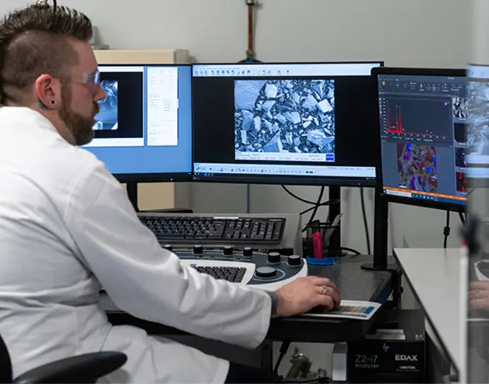
A chance find in the field became an opportunity to characterise the wing’s micro- and nanoscale structure, and here we use it to demonstrate how correlative microscopy links complementary imaging techniques to reveal detail that no single method could capture alone.
Our approach
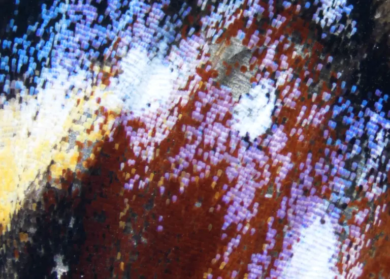
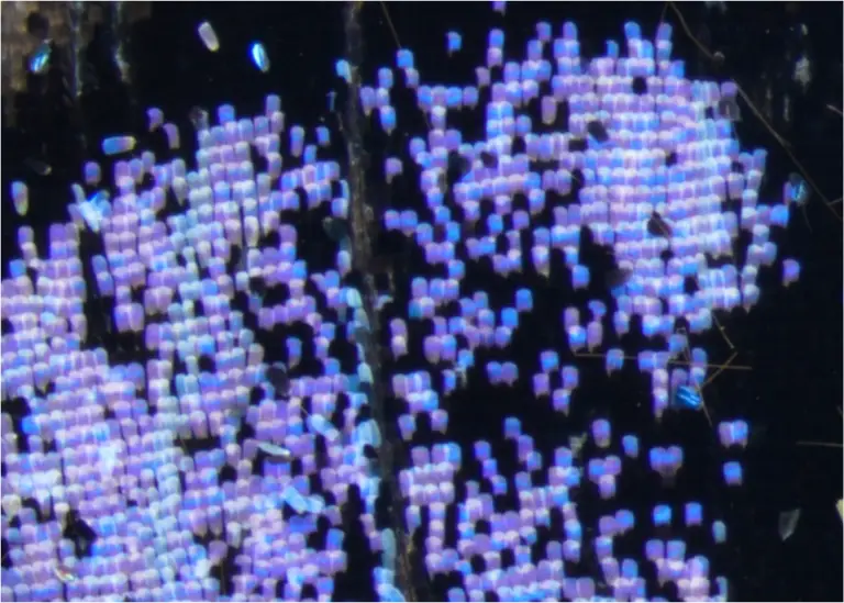
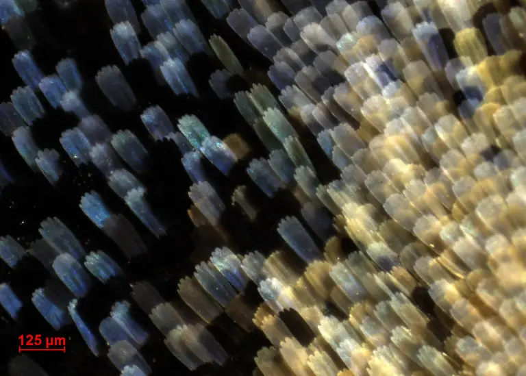
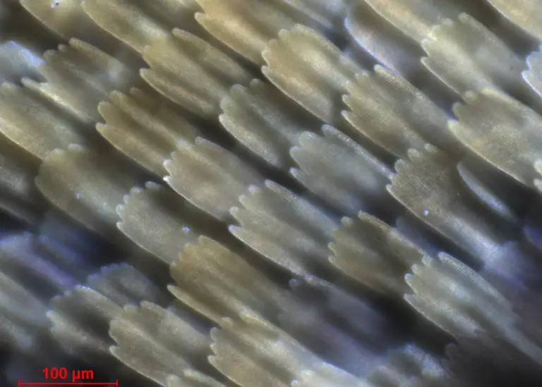
Light microscopy images were collected using a Nikon SMZ745-T Stereo Light Microscope and a Nikon Eclipse Ni-U Compound Light Microscope.
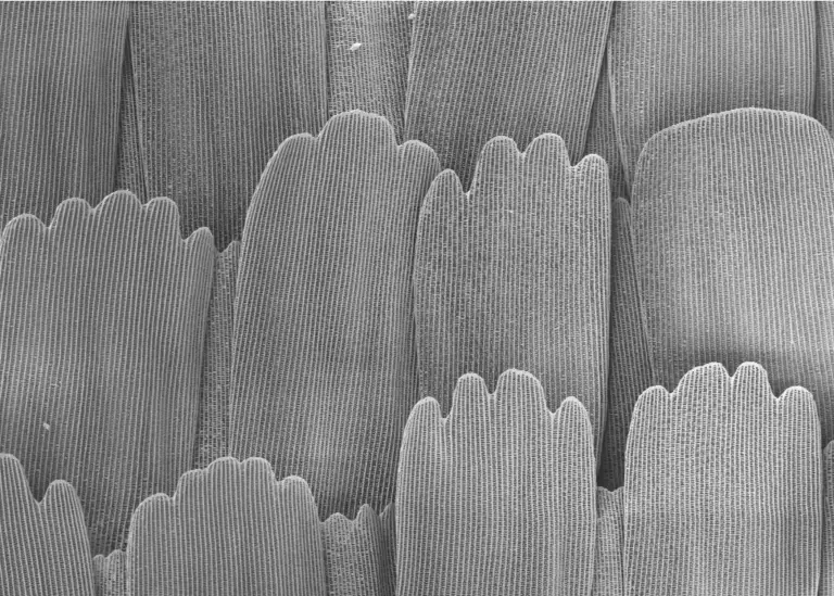
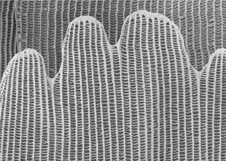
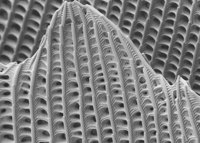
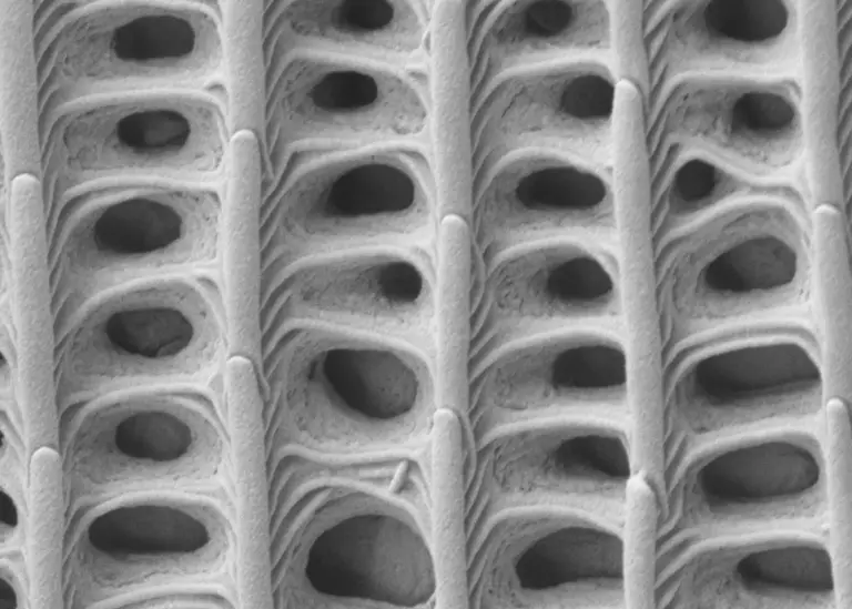
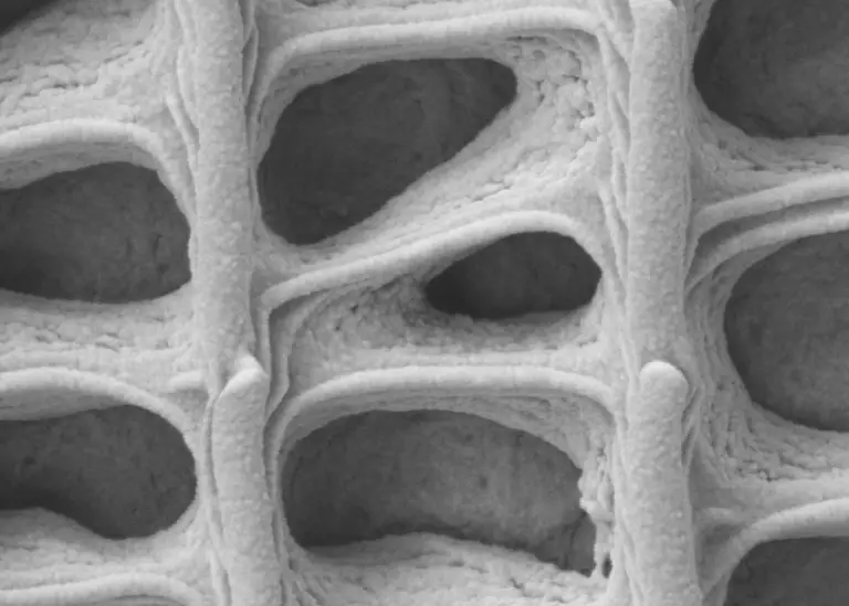
Electronmicrographs were generated using a Zeiss Sigma Field Emission SEM with a secondary electron (SE2) detector at 3 kV accelerating voltage with 30 µm aperture.
Results
Taken together, the image series follows the butterfly wing across multiple length scales, from whole-wing architecture and colour patterning observed by light microscopy to the fine surface features and textures revealed by SEM. As magnification increases, the organisation of overlapping scales becomes apparent, followed by increasingly detailed views of the ridges, cross-ribs and lamellar structures that define the wing surface. These features influence how light interacts with the scales and contribute to the wing’s distinctive appearance.
What this means
The study demonstrates the value of a correlative microscopy approach. Light microscopy provides the broader context, showing overall morphology, colour distribution and scale arrangement, while SEM reveals the surface details needed to understand structural relationships at the micro- and nanoscale. Viewed together, these techniques provide a more complete picture than either method alone.
While a butterfly wing provides a visually engaging example, the same workflow is routinely applied to industrial and scientific investigations. Whether examining particles, fibres, coatings, contaminants, surface defects, material interfaces or product morphology, correlative microscopy helps connect what is observed at the macroscopic level with the underlying structures responsible for it and their interactions. This combination of context and high-resolution evidence supports stronger interpretation and more confident decision-making across research, development, troubleshooting and characterisation projects.
Have a feature, particle, or surface you can’t explain?
Structural detail that is invisible to one technique is often obvious to another. Resolian’s microscopy team pairs light microscopy with SEM and EDX, matching the technique and detector mode to the question in front of you, so you can move from a visible problem to a resolved one with clear, defensible interpretation behind it.