Microscopy and Imaging Services
Light Microscopy & Scanning Electron Microscopy (SEM) & Dynamic Image Analysis (QICPIC)
Imaging insights for material characterization and investigation
cGMP
LABORATORIES
20+ YEARS
ANALYTICAL EXPERTISE
FDA & MHRA
AUDITED
Microscopy and Imaging techniques are widely used to characterize materials, particles, surfaces, crystalline purity and product components. Whether supporting routine material assessment, crystallization, formulation development, manufacturing investigations, or contamination studies, microscopy provides visual and structural information that helps build a clearer understanding of your sample.
At Resolian, these techniques are complementarily used with analytical techniques as part of a broader characterization strategy, helping clients answer practical questions about material properties, composition, performance and drive the project forward.
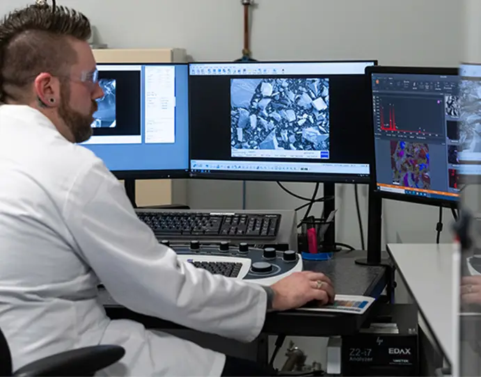
When the answer matters
Resolian’s microscopy and imaging capabilities support the characterization of pharmaceutical materials, products, packaging components, and isolated particles throughout the project lifecycle, and for quality investigations.
Our analysts combine microscopy expertise with a broader understanding of pharmaceutical development and analytical sciences, delivering representative high-quality output, scientifically grounded observations, and data that can be integrated into wider project decisions.
Our integrated capability
We select the appropriate microscopy approach based on the sample type, study objectives, the information provided and the outcome required. Our capability includes light microscopy, SEM imaging, SEM-EDX elemental analysis, hot-stage microscopy and dynamic image analysis, which can be applied individually or as part of a wider analytical programme.
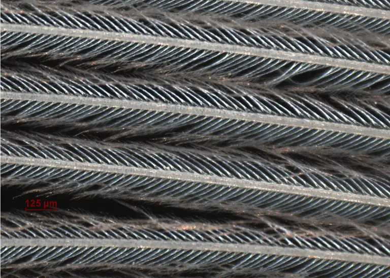
Light Microscopy
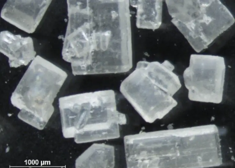
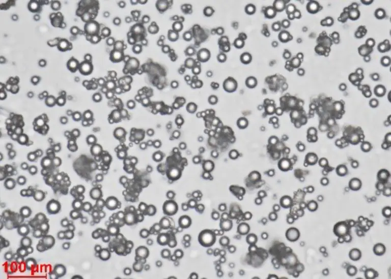
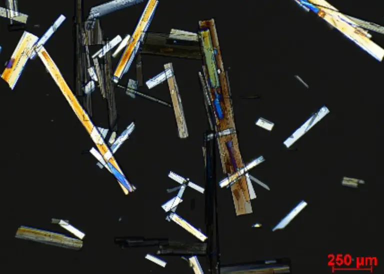
Light microscopy is frequently the starting point for material and particle characterization. It provides information on morphology, color, size, optical properties, crystallinity and purity, as well as sample heterogeneity, supporting both routine assessments and more detailed investigations.
In some cases, light microscopy alone provides sufficient information. Where additional surface detail or compositional information is required, further investigation using SEM or SEM-EDX may be recommended.
Our suite of Stereo and Compound microscopes enable a broad range of characterization approaches, including:
- Reflective Light Microscopy
- Bright-Field Microscopy
- Dark Field Microscopy
- Differential Interference Contrast (DIC) Microscopy
- Polarized Light Microscopy
- Hot Stage Microscopy
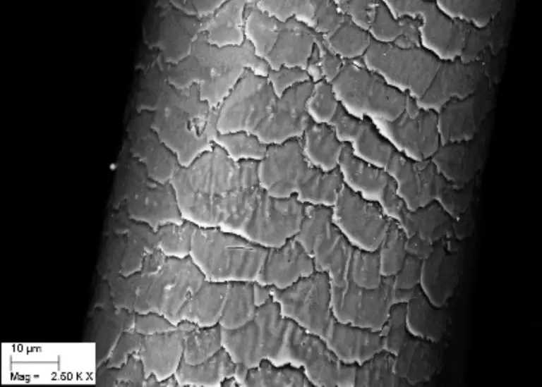
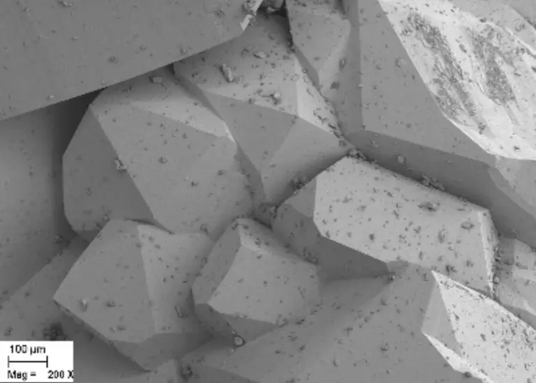
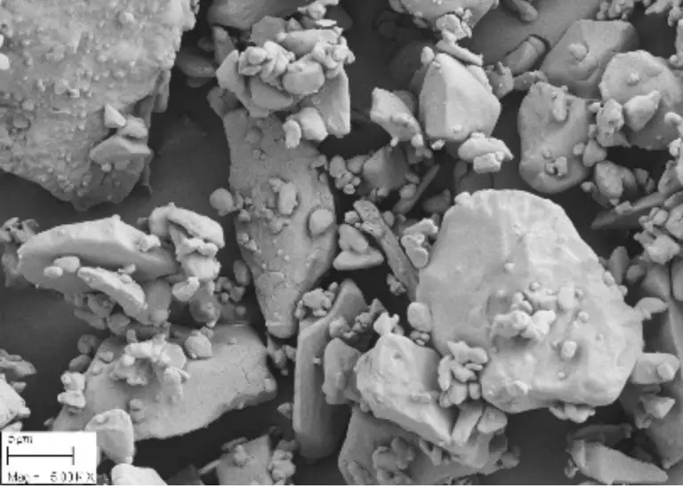
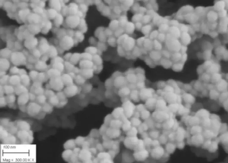
Scanning Electron Microscopy (SEM)
Reveal detail beyond the limits of light microscopy with SEM services that deliver higher-resolution surface imaging, improved depth of field, and clearer particle morphology assessment.
Our SEM imaging can be tailored using different detector modes, allowing you to select the most appropriate contrast, surface detail, or compositional information for the investigation.
- Secondary electron detector: ideal for high-detail surface topography, fine morphology, and texture assessment, helping visualise particle shape and texture.
- Backscattered electron detector: highlights compositional contrast, making it useful for distinguishing phases, inclusions, coatings, and particles with different average atomic composition.
- In-lens detector: provides enhanced surface sensitivity and crisp imaging of very fine features, supporting high-resolution assessment of delicate or low-relief material surfaces.
Different detector modes provide complementary information, allowing our scientists to select the most appropriate imaging approach for the analytical question being addressed.
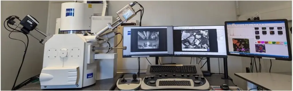
Energy Dispersive X-ray (EDX)
Our system is equipped with energy dispersive X-ray probes for elemental analysis. EDX analysis identifies the elemental composition of selected particles, residues, inclusions, or surface features, adding chemical context to SEM observations.
By comparing elemental profiles across suspect and reference materials, EDX can help distinguish inorganic contaminants, support source investigations, and guide next-step testing. It is particularly useful when visual appearance alone can’t confirm whether a particle is formulation-related, process-derived, environmental, or extraneous.
Elemental analysis can provide important contextual information alongside microscopy observations, helping to differentiate materials and support further scientific assessment.
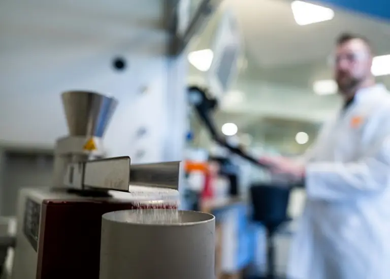
Dynamic Imaging Analysis
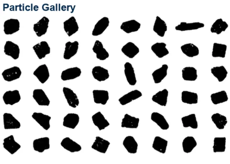
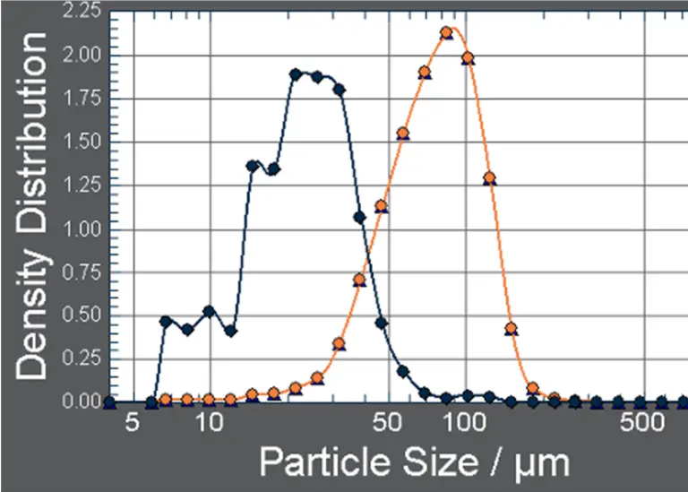
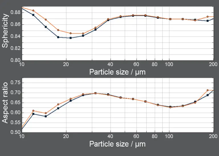
Dynamic image analysis provides quantifiable particle size and shape information in a single, image-based measurement, helping pharmaceutical teams understand material attributes that can influence formulation its performance, manufacturability and product robustness.
Using the Sympatec QICPIC platform, Resolian can assess APIs, excipients, intermediates and finished products, measuring particle size and various shape descriptors. This supports formulation development, particle engineering, milling optimization, granulation assessment, troubleshooting, batch-to-batch comparison and contamination.
The modular QICPIC platform allows dispersion conditions to be matched to the material and analytical objective, with fast dry dispersion for cohesive powders, gentler gravity-based options for fragile, coarse or free-flowing materials, and wet dispersion for suspensions, emulsions or samples best characterized in liquid media.
With wet or dry dispersion, statistically meaningful results can be reported as clear distributions, comparative datasets and particle image evidence, giving practical insight into how material attributes relate to performance and processability.
Underpinned by expert interpretation
Microscopy results are reviewed and interpreted by experienced analysts, ensuring that observations are reported within the context of the study objectives and the wider analytical programme..
For a full contamination investigation combining these techniques with FTIR, PXRD and mass spectrometry against USP and Ph. Eur. standards, see our Foreign Matter Analysis service.
Discuss your requirements with our scientific team.
We can help determine the most appropriate analytical approach for your material characterization or investigation project.
Proven in practice
Real investigations our microscopy team has helped resolve.
From a Butterfly Wing to Nanoscale Structure: What Correlative Microscopy Reveals
Why choose Resolian for microscopy services?
- Light microscopy, SEM, and SEM-EDX capabilities within a broader analytical sciences offering
- Scientists who combine microscopy expertise with pharmaceutical development insight
- cGMP laboratories, audited by the FDA and MHRA
- Scientifically interpreted reports tailored to study objectives
- Microscopy data that can be integrated with complementary analytical techniques when required
Your Goals. Our Science.
Talk to our team
From routine material characterization to targeted investigations, our microscopy team provides the imaging and scientific interpretation needed to support your analytical objectives.
Our Analytical Sciences Resources

Case Studies
From a Butterfly Wing to Nanoscale Structure: What Correlative Microscopy Reveals


Frequently Asked Questions
Find answers to commonly asked questions about light microscopy and SEM at Resolian.
What's the difference between light microscopy and SEM?
Light microscopy gives a fast first look at a particle or surface, color, shape, and behavior under polarized or reflected light. SEM goes further, resolving detail beyond the limits of light and, with EDX, adding elemental composition.
When do I need SEM instead of light microscopy?
When a feature is too fine for light to resolve, or when you need to confirm composition rather than just appearance.
What does EDX add to an SEM image?
EDX identifies the elemental composition of a selected particle or feature, adding chemical context that helps confirm whether something is formulation-related, process-derived, or extraneous.
Which SEM detector mode is right for my sample?
It depends on the question. Secondary electron detection suits fine surface texture, backscattered electron detection highlights compositional contrast, and in-lens detection suits delicate, low-relief surfaces. Our analysts select the mode for your investigation.
How is this different from Resolian's Foreign Matter Analysis service?
This page covers our core light and electron microscopy capability. Foreign Matter Analysis applies that capability, alongside FTIR, PXRD, and other techniques, to full contamination investigations against USP and Ph. Eur. standards.
What sample types can you examine?
Raw materials, APIs, excipients, formulated products, packaging components, and isolated particles or residues.
What do I receive at the end of an investigation?
Representative images, clear observations, and a written scientific interpretation, not just a folder of images.